
QT Imaging utilizes transmission and reflection acoustic energy to generate highly detailed images of the human anatomy. The QTI Breast Acoustic CTTM System is FDA-cleared for breast imaging. QT Imaging team seeks to adapt the technology platform may one day offer whole body applications including prostate cancer screening, orthopedics, infant imaging, and full body screening.
The QT Imaging team is pleased to share many of the exciting projects our research team is working on. The products and technology described in this section are not all FDA-cleared.
Fibroglandular ratio measurement is an innovative technology that promises to measure breast density, with no radiation or compression. This feature is FDA cleared to provide FGV measurement as well as ratio of FGV to TBV, which is the surrogate measurement of breast density.
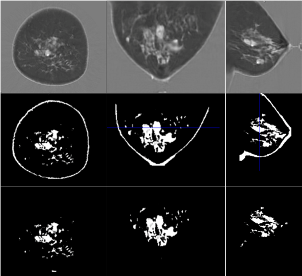
Steps in the quantitative breast density algorithm:
QT Imaging’s research has the potential to measure and follow masses in the breast with high accuracy and precision, putting this technology in the same category with CT and MRI, rather than the hand-held ultrasound. Our technology generates 3D speed-of-sound maps that can be used to perform volumetric measurement and segmentation of breast tissue. Tumor volume doubling time is associated with growth rate and tumor biology. The QTI team is working on providing the accurate measurement of tumor volume change-over-time which is critical for oncologic diagnosis, staging, and treatment, including quantification of neoadjuvant response.
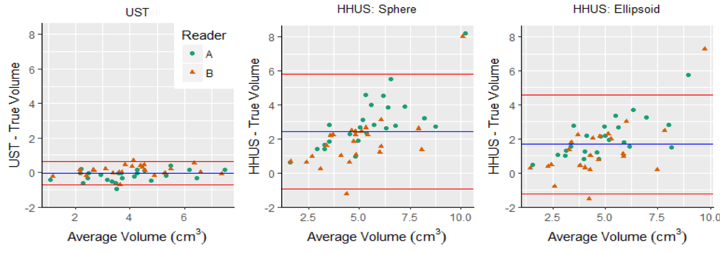
Bland-Altman plots showing absolute agreement between volume of mass assessed using (left) ground truth and QT, (middle) ground truth and Hand-Held Ultrasound (HHUS)-spherical method, and (right) ground truth and HHUS-ellipsoid method. The spherical and ellipsoid methods corresponding to volume calculation formulas differ in that the spherical method uses the average of the three ellipsoidal radii in each orthogonal direction. The ellipsoidal method uses the three ellipsoidal diameters directly.
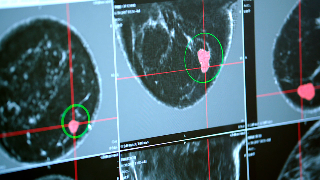
QT Imaging’s research and development includes what may soon be the first artificial intelligence-enabled algorithm using transmission and reflection acoustic energy modalities to aid in the diagnosis of cancer. These modalities supply quantitative (objective) measures at resolution previously not possible. The reflection mode is compounded over 360 degrees and avoids artifacts by utilizing the speed of sound to correct for refraction and attenuation to guide gain. This removes speckle, making this modality more amenable for use in AI/ML training.
The speed of sound, attenuation, and reflection images that minimize artifacts, by leveraging 3D modelling of the acoustic energy in the breast.
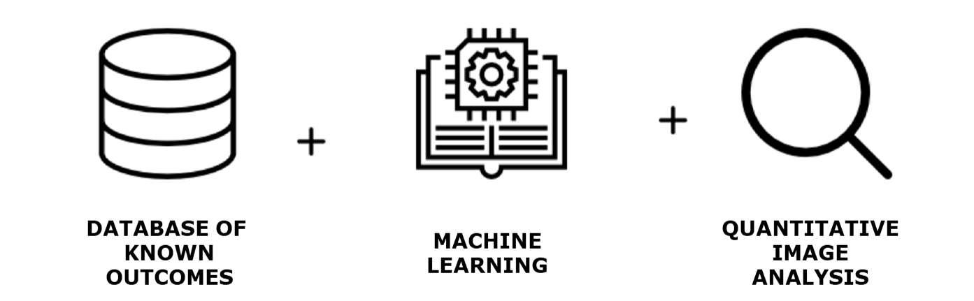
AI/ML-based algorithms have gained tremendous acceptance throughout medical imaging community. QT Imaging team strives to provide radiologists with a tool that can increase image specificity. QTviewer® with additional AI/ML tools may enable features analysis to categorize findings as either benign or malignant. Our goal is to markedly reduce false positives and unnecessary biopsies. QT Imaging is uniquely positioned to accomplish this since it has amassed a unique library of ~15,000 breast images from installations in the US (multiple states), and elsewhere. This is not an FDA cleared feature just yet.
QT Imaging is also working on implementing a machine learning/convolutional neural network (ML/CNN) based denoising method that removes artifacts in the water and breast without compromising diagnostic capability. ML will also be utilized for image formation and data cleansing.
The QT Imagine 3D ultrasound tomography algorithm has been shown to be mathematically equivalent to training a large convolutional deep neural network with certain symmetry. This explains the near optimal performance of the algorithm on various GPU architectures.
The QTI Breast Acoustic CT™ System is FDA cleared for breast imaging and is a near isotropic 3D transmission/reflection acoustic energy technology without harmful radiation or painful compression. The technology produces high-fidelity detailed images, providing clarity even for women with dense breasts.
3D ultrasound tomography means that the artifacts are minimized by modeling acoustic energy in full 3D and collecting 3D data on a 2048 element array. The computationally intensive full 3D modelling reconstruction is made possible by suitable mathematics and use of GPUs.
Looking forward, these new imaging modalities have the potential for additional clinical benefits as QT Imaging’s transmission/reflection acoustic computed tomography has imaged, independently verified, microanatomy in living humans and has identified human tissue types using speed of sound, as part of QT Imaging’s research.
The QT Imaging 3D technology delivered the first ever 3D printed human breast tissue-specific microanatomy using any medical imaging modality and has the potential to be a new diagnostic tool, and a new way for physicians to visualize human health and disease.
QT Imaging’s 3D Acoustic Computed Tomography offers the potential to be a new diagnostic imaging modality for the whole body, including the most difficult regions where bone and air are present.
The QT Imaging team has verified the accuracy of measured tissue speed-of-sound values a potential biomarker even in the presence of bone and air and provided evidence of the ability to utilize the second slow compressional wave uniquely present in the bone’s porous medium (trabecular bone).
QT Imaging’s technology may provide:
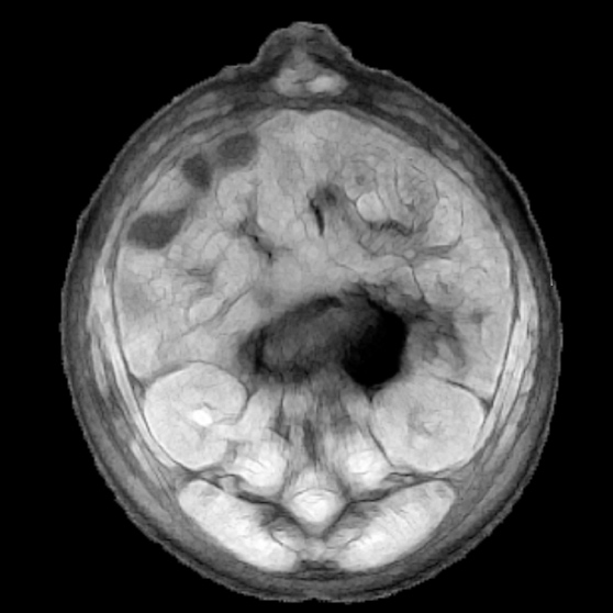
QTscan®
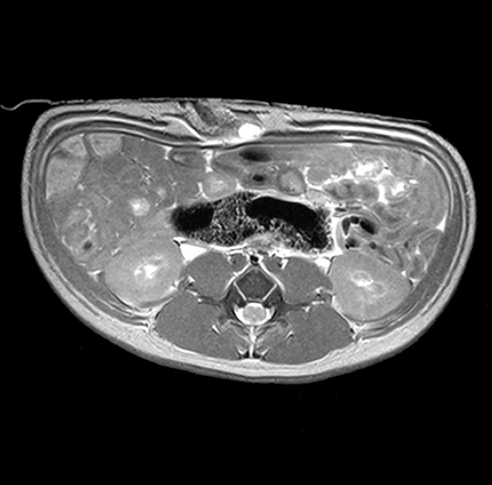
MRI
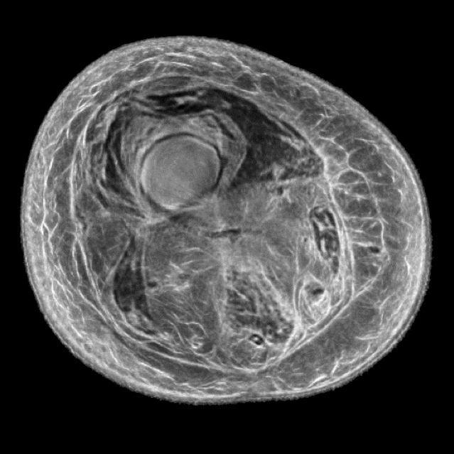
QTscan®
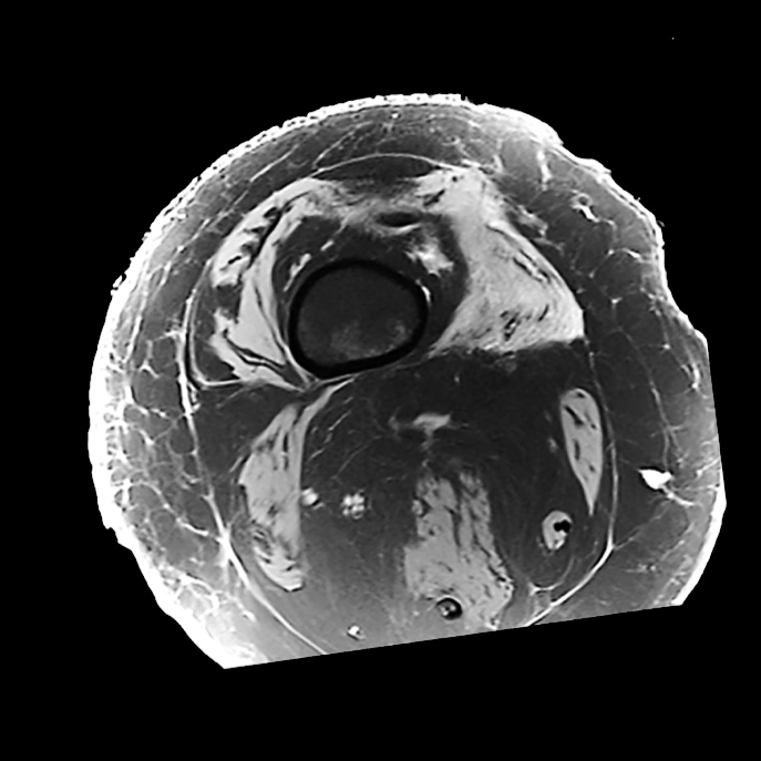
MRI
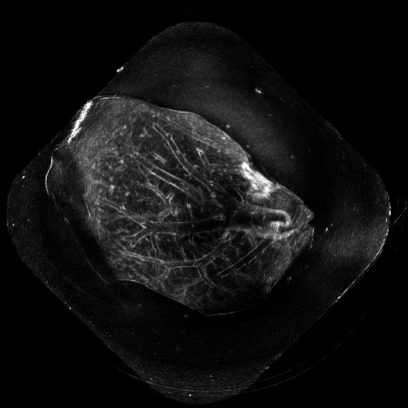
QT reflection image of a liver
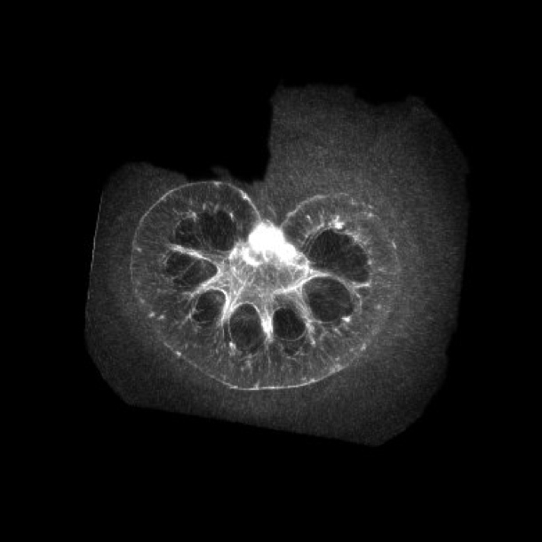
QT reflection image of a kidney