Breast cancer is the most commonly diagnosed women’s cancer in the United States, according to the National Cancer Institute. The American Cancer Society estimates that in 2022, 287,850 women in the United States have been diagnosed with invasive and in situ (early stage) breast cancer, and breast cancer has claimed the lives of 40,920 women. 1 The American Cancer Society further estimates that one out of every eight women will develop breast cancer at some point during her life and one in every 42 women who turns 50 today will have a diagnosis of breast cancer before she turns 60.
There are several dominant screening and diagnostic technologies that are used both independently and dependently to locate cancers at an early stage and improve treatment outcomes. Each of the currently available non-surgical modalities for breast cancer detection has various clinical limitations. Screening methods and technologies include: (i) breast self-examination and clinical breast examination; (ii) mammography, including screening mammography, diagnostic mammography, and mammography with computer aided detection; (iii) handheld ultrasound (HHUS) and (iv) magnetic resonance imaging (MRI).
Mammography is the dominant imaging modality in today’s standard of care. The American Cancer Society recommends that women of average risk have the option to begin mammography at age 40, get mammograms every year from age 45-54, and have mammography every other year starting at age 55. 2 Despite those recommendations, only 65% of women over 40 in the United States have had a mammogram in the previous two years, and only 58% of women between 40 and 49 had a mammogram in the previous two years; 3 even though, screening mammography is 100% covered under the Affordable Care Act. This is in part due to the limitations of mammography, both in terms of sensitivity and reliability for dense breast tissue, where 10-15% of cases have inconclusive results requiring further testing, as well as concerns about safety.
Mammography is also problematic in women who have breast implants. For these women, problems include painful mammograms, delayed detection of cancer from interference in imaging breast tissue, and an unwillingness to perform mammograms due to fear of implant rupture, dislocation or capsular contracture.
QT Imaging’s goal is to provide highly accurate, 100% safe and painless breast imaging that can be used to:


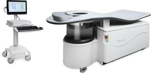

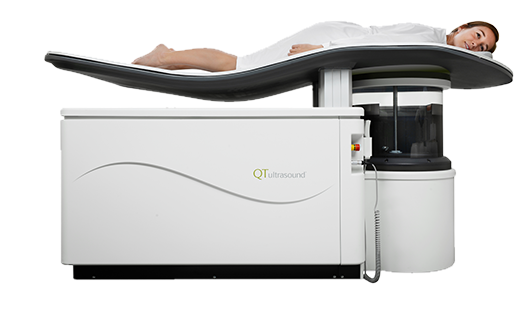
The Breast Acoustic CT™ system is a non-invasive, FDA cleared, breast imaging tool. Transmission ultrasound together with reflection ultrasound provide highly complementary and synergistic information about breast tissue, which can enable healthcare providers to effectively assess breast health. This evolution in medical imaging technology generates 3D images with remarkable accuracy and precision, and the volumetric images are produced without the need of contrast agents or radiation.


What makes this emerging technology important is that the QTscan™ transmission imaging is quantitative in nature, and thus provides us with information which can assist clinicians in identifying different breast tissues.
In a QTscan™, images are generated using both reflection and transmission modalities. QT Imaging’s technology developments in hardware and imaging algorithms have enabled improvements in visualizing breast tissues in a natural pendant state.
The Breast Acoustic CT™ system uses a transmitter/receiver array pair as well as a system of reflection transceivers to create a multi-modality system (transmission and reflection). Multiple transmission acquisitions are acquired for 180 angles as the transmitter is rotated fully around the subject. Reflection data from the reflection transducers is interleaved between the transmission acquisitions. The transmission and reflection images align with each other, thus increasing diagnostic capability.
Because this innovative model-based imaging technology utilizes true 3D data acquisition and reconstruction, QT Imaging’s image reconstruction algorithm can systematically characterize and isolate different tissue types based on speed of sound and other tissue properties.
 QT information visually correlates with histology

QT information visually correlates with histology
John C. Klock et al.
International Journal of Biomedical Imaging Volume 2016, Article ID 7570406, 9 pages
A QT Imaging system uses a transmitter/receiver array pair as well as a system of reflection transceivers to create a multi-modality system (transmission and reflection). Multiple transmission acquisitions are acquired for 180 angles as the transmitter is rotated fully around the subject. Reflection data from the reflection transducers is interleaved between the transmission acquisitions. The result is a 3D speed-of-sound (SOS) image and reflection tomogram.
Because this innovative imaging technology utilizes true 3D data acquisition and reconstruction, the QT algorithm is able to systematically characterize and isolate certain tissues based on speed of sound.
Conventional breast imaging modalities, such as mammography, face tremendous challenges when imaging dense breast tissue, which puts women with dense breasts – nearly half the female population – at an immediate disadvantage and at risk of misdiagnosis. This is because cancer can appear similar to regular dense breast tissue and if there is a lot of dense breast tissue, the cancer can ”hide.” QT Imaging technology has the ability to detect cancer within dense breasts, and our ongoing clinical trials aim to confirm how our Breast Acoustic CT™ system can effectively and consistently identify suspicious regions. If no cancer is identified, confidence is high that the breast is healthy. QTscan™ shows high contrast breast images with no need for injections with contrast agents and no exposure to radiation.
Mammography

Mammography
3D Mammography with Tomosynthesis
Handheld Ultrasound

Handheld Ultrasound
Breast MRI

Breast MRI
QTscan

QTscan
Case study of single subject with suspicious mass as viewed across multiple imaging modalities.
Mammography
(not visible)

Mammography
(not visible)
Handheld Ultrasound

Handheld Ultrasound
Breast MRI
(non-enhancing)

Breast MRI
(non-enhancing)
QTscan

QTscan
Case study of Invasive Lobular Carcinoma as viewed across multiple imaging modalities.
See the rest of the comparative case studies Read our Dense Breast Mass Detection Study
The QTscan’s true 3D acquisition and 3D reconstruction offers a new way to look at breast imaging and breast health. See for yourself.